Anterior Pituitary adenohypophysis creates a number of different hormones. Terminologies for the components of the pituitary gland are based on the embryological origins of the main subdivisions as well as the anatomical regions of each.
Anatomy A215 Virtual Microscopy
The pituitary endocrine gland which is located in the bony sella turcica is attached to the base of the brain and has a unique connection with the hypothalamus.

. PITUITARY HORMONES PITUITARY GLAND HYPOPHYSIS DIAMETER. Between these lobes lies a small. The anterior pituitary synthesizes and secretes somatotropin prolactin follicle stimulating hormone luteinizing hormone thyroid hormone adrenocorticotropic hormone.
It arises from two different tissue sources. The basophils appear as darker cells with purple cytoplasm. A anterior lobe b posterior lobe.
Pituitary glands and the hypothalamus together act as master regulators of the endocrine system. The location of the anterior and posterior pituitary is the most obvious difference between them. -Supraoptic nucleus and paraventricular nucleus are origin of posterior lobe of pituitary.
Slide of Pituitary Gland In the space below draw and label the anterior pituitary gland and the posterior pituitary gland. The posterior pituitary is composed of neural tissue. Submandibular or Digastric Triangle Boundaries.
This gland is located just below the brain somewhat behind the eyes. The hormones secreted by. The anterior pituitary gland is a part of the pituitary at the base of the brain and produces hormones for the regulation of physiological functions such as growth reproduction lactation and stress.
System of glands that secrete hormones directly into the blood. In clinical reference the term anterior pituitary is often used synonymously with pars distalis and posterior pituitary is frequently used synonymously with pars nervosa. Chromophils- stain with HE and secrete hormones.
The endocrine system is the only system where t. A What hormones from the hypothalamus will. Dont forget to join social media of anatomy learner for more update pituitary gland histology labeled pictures and diagram.
These cells are mammotrophs and somatotrophs. Divides into to lobes. Make a sketch of your observed field of view and label anterior.
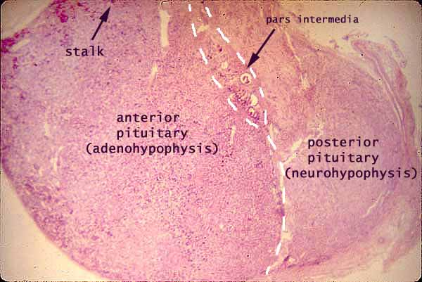
Congenital pituitary hormone deficiencies have been reported in approximately one in 4000 live births however studies reporting mutations in some widely studied transcription factors account for only a fraction of congenital hormone deficiencies in. It is formed from a downgrowth of the diencephalon that forms the floor of the third ventricle. Switch to low power and observe the area that separates the two parts as shown in Figure 113.
This slide shows a section of the human pituitary. Anterior Pituitary vs Posterior Pituitary. Posterior pituitary is nervous tissue neurohypophysis and anterior pituitary is glandular adenohypophysis.
The anterior part is derived from an upgrowth from the oral ectoderm of the primitive oral cavity called Rathkes pouch. The anterior pituitary the posterior pituitary. Anatomy Test 4 Endocrine System slides endocrine system.
In the anterior pituitary pars distalis you can see cords of cuboidal cells with a wide range of nuclear to cytoplasmic volume ratios. Looking at it under a microscope and it looks busy It contains a large number of cells called secretory cells. This is the view under a dissecting microscope.
The latter structure may be better seen on slide 122. PITUITARY GLAND Has two parts. The pituitary gland sometimes called the hypophysis is a small gland that dangles from the base of the brain like a pea on a string Several hormones produced by the hypothalamus are stored here and released into the blood.
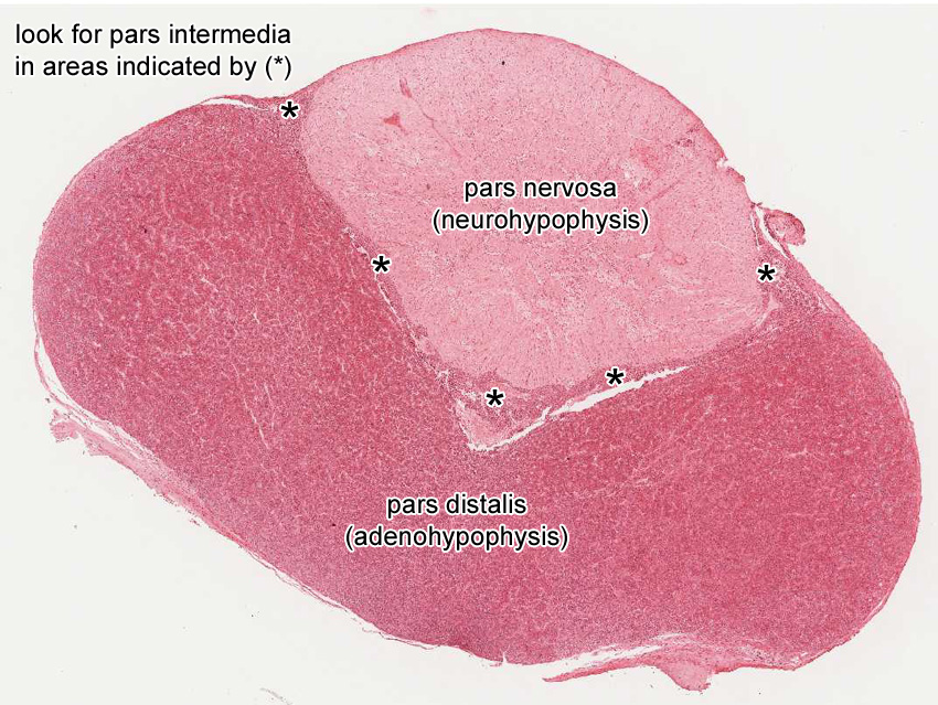
Along the posterior part of the the anterior lobe there is a narrow region called. HORMONES OF ANTERIOR PITUITARY GROWTH HORMONE. Pars Distalis - comprises most of the anterior lobe 75 and contains five types of endocrine cells.
The posterior pituitary does not synthesize hormones it stores and releases vasopressin and oxytocin. Submandibular gland duct CN XII Nerve to mylohyoid Submental artery Submental lymph nodes Suprahyoid muscles. The posterior pituitary pars nervosa is connected to the hypothalamus by the pituitary stalk - this is easily visualized because the nervous tissue appears continuous between the two glands.
Anterior and posterior pituitary hormones. Pituitary gland weighs approximately 500mg to 900mg and lies immediately beneath the third ventricle and just above the sphenoidal sinus in the sella turcica Turkish saddle. How can you tell the difference between anterior and posterior pituitary.
Difference Between Anterior and Posterior Pituitary Gland Definition. Lateral Anterior posterior bellies of Digastric muscle 1 Superior Inferior border of mandible 2 Floor Mylohyoid muscle 3 Hyoglossus muscle and Middle constrictor of pharynx 1 2 3 Contents. Finally a few chromophobes are visible in this.
The acidophils appear as cells with pink cytoplasm and dark nuclei. -Connected to hypothalamus via hypothalamohypophyseal tract. Posterior Pituitary neurohypophysis does not create hormones it stores hormones from the hypothalamus.
Posterior pituitary-is the neural portion derived from an extension of the hypothalamus median eminence which remains connected throughout life by a stalk called the infundibulum Fig 1. Obtain a prepared slide of the pituitary gland Begin observing it under scanning power and notice the difference between the anterior pituitary and posterior pituitary tissue organization. As the human embryo develops the anterior pituitary is formed from cells from the roof of the mouth that migrate toward the brain.
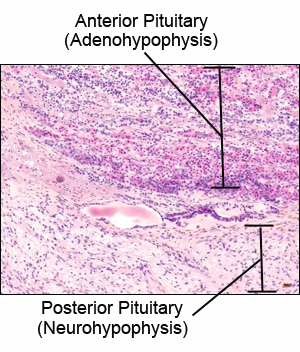
The anterior lobe adenohypophysis stains darker and the posterior lobe neurohypophysis stains lighter. The pituitary gland is composed of an anterior and posterior lobes. These are the corticotrophs thyrotrophs and gonadotrophs.
The pituitary gland consists of two anatomically and functionally distinct regions the anterior lobe adenohypophysis and the posterior lobe neurohypophysis. Examine a section of pituitary slide 128 103 and identify the anterior pituitary the posterior pituitary and the intervening pars intermedia with cysts representing remnants of Rathkes pouch Figs. The pars intermedia is poorly developed in humans.
The pituitary gland is split into two different portions and the anterior is at the front while the posterior is in the back. Supporting cells called pituicytes make up about one fourth or 25 of. The normal microscopic appearance of the pituitary gland is shown above.
Anterior Pituitary- contains three divisions. This slide displays the three cell types of the anterior pituitary under HE stain. Hope this guide was helpful to learn pituitary gland histology.
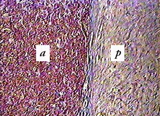
The anterior and posterior pituitary hormones and their target organs. While the anterior pituitary a is made up almost entirely of cells the posterior pituitary p contains few cells and a lot of nerve cell processes--the axons of hypothalamic neurons. SELLA TURCIA TWO PORTIONS 1- ANTERIOR PITUITARY ADENOHYPOPHYSIS 2-POSTERIOR PITUITARY NEUROHYPOPHYSIS 2.
The posterior part of the pituitary has its embryological origins in nervous tissue. Draw in the location of the hypothalamus and pineal gland with respect to pituitary gland. The posterior pituitary gland is an endocrine.
Now you will able to identify the anterior and posterior pituitary gland histology slide under microscope with important identification points. -Posterior pituitary neurohypophysis consists of axons from hypothalamic nuclei and glial cells.

A P Ii Endocrine Gland Slides Flashcards Quizlet

Endocrine Systems Lab
Endocrine System Histology

Endocrine Systems Lab

Endocrine Systems Lab

Pin On Histology A P Ii
Histology At Siu
Pituitary Gland
0 comments
Post a Comment